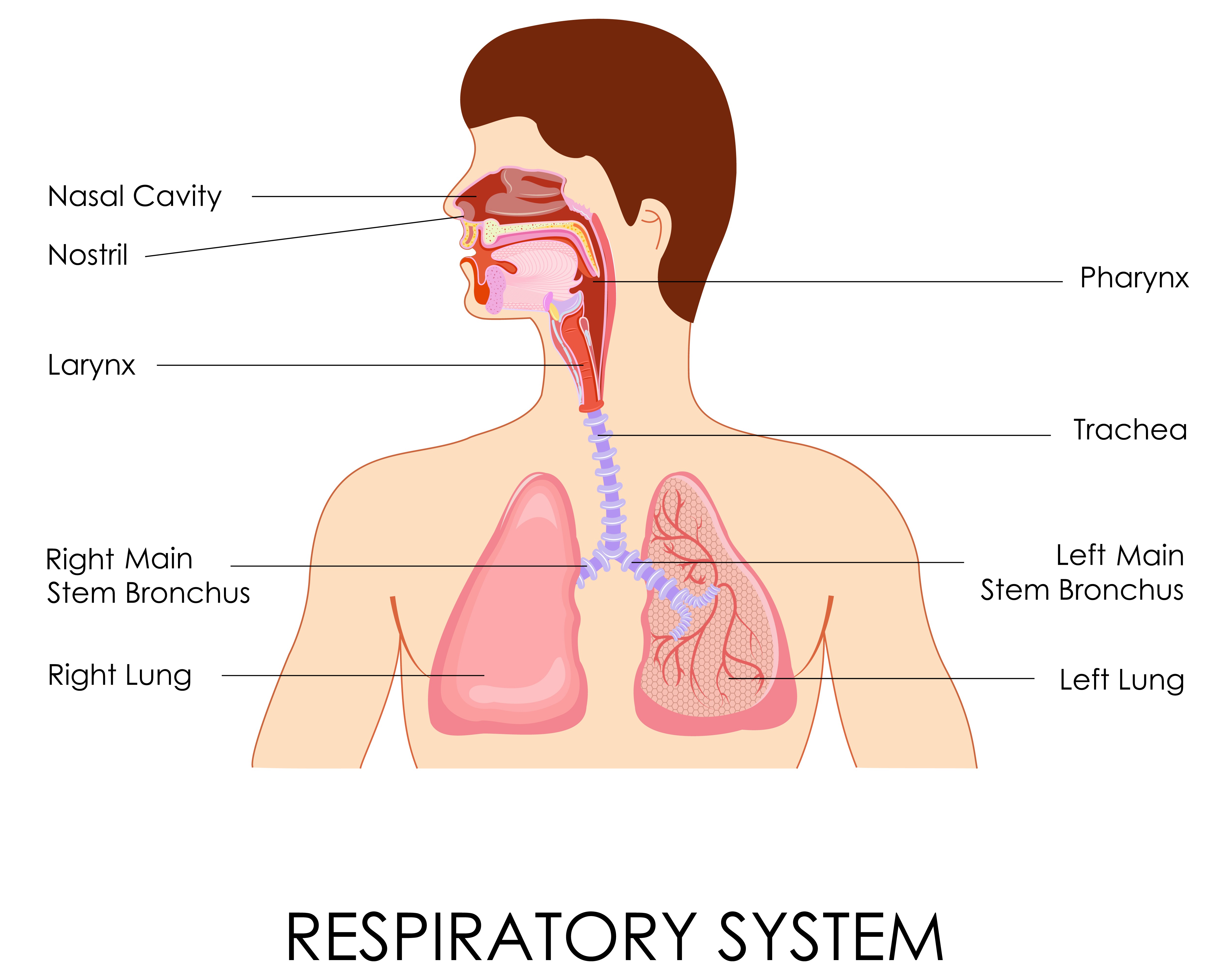
Pulmonary Oedema

5 stars based on 38 reviews

Pulmonary edema is fluid accumulation in the tissue liquids and gases are both fluids in lungs air spaces of the lungs. It is due to either failure of the left ventricle of the heart to remove blood adequately from the pulmonary circulation cardiogenic pulmonary edemaor an injury to the lung parenchyma or vasculature of the lung noncardiogenic pulmonary edema. Pulmonary edema, especially acute, can lead to fatal respiratory distress or cardiac arrest due to hypoxia. It is a cardinal feature of congestive heart failure.

The most common symptom of pulmonary edema is difficulty breathingbut may include other symptoms such as coughing up blood classically seen as pink, frothy sputumexcessive sweatinganxietyand pale skin. These are liquids and gases are both fluids in lungs presenting symptoms of chronic pulmonary edema due to left ventricular failure.

The development of pulmonary edema may be associated with symptoms and signs of "fluid overload"; this is a non-specific term to describe the manifestations of right ventricular failure on the rest of the body and includes peripheral edema swelling of the legs, in general, of the "pitting" variety, wherein the skin is slow to return to normal when pressed uponraised jugular venous pressure and hepatomegalywhere the liver is enlarged and may be tender or even pulsatile.

Other signs include end-inspiratory crackles sounds heard at the end of a deep breath on auscultation and the presence of a third heart sound. Classically it is cardiogenic left ventricular but fluid may also accumulate due to damage to the lung. This damage may be direct injury or injury mediated by high pressures within the pulmonary circulation.

When directly or indirectly caused by increased left ventricular pressure pulmonary edema may form when mean pulmonary pressure rises from the normal of 15 mmHg [3] to above 25 mmHg. By convention cardiogenic refers to left ventricular causes.

Injury to the lung may also cause pulmonary edema through injury to the vasculature and parenchyma of the lung. Some causes of pulmonary edema are less well characterised and arguably represent specific instances of the broader classifications above. Flash pulmonary edema FPEis rapid onset pulmonary edema. It is most often precipitated by acute myocardial liquids and gases are both fluids in lungs or mitral regurgitationbut can be caused by aortic regurgitationheart failureor almost any cause of elevated left ventricular filling pressures.

Treatment of FPE should be directed at the underlying cause, but the mainstays are ensuring adequate oxygenation, diuresisand decrease of pulmonary circulation pressures. Recurrence of FPE is thought to be associated with hypertension [15] and may signify renal artery stenosis.

There is no one single test for confirming that breathlessness is caused by pulmonary edema; indeed, in many cases, the cause of shortness of breath is probably multifactorial. Low oxygen saturation and disturbed arterial blood gas readings support the proposed diagnosis by suggesting a pulmonary shunt. Chest X-ray will show fluid in the alveolar walls, Kerley B linesincreased vascular shadowing in a classical batwing peri- hilum pattern, upper lobe diversion increased blood flow to the superior parts of the lungand possibly pleural effusions.

In contrast, patchy alveolar infiltrates are more typically associated with noncardiogenic edema [2]. Lung ultrasound, employed by a healthcare provider at the point of care, is also a useful tool to diagnose pulmonary edema; not only is it accurate, but it may quantify the degree of lung water, track changes over time, and differentiate between cardiogenic and non-cardiogenic edema.

Especially in the case of cardiogenic pulmonary edema, urgent echocardiography may strengthen the diagnosis by demonstrating impaired left ventricular function, high central venous pressures and high pulmonary artery pressures. Blood tests are performed for electrolytes sodium, potassium and markers of renal function creatinine, urea.

Liver enzymesinflammatory markers usually C-reactive protein and a complete blood count as well as coagulation studies PT, aPTT are also typically requested. B-type natriuretic peptide BNP is available in many hospitals, sometimes even as a point-of-care test. Liquids and gases are both fluids in lungs those with underlying heart disease, effective control of congestive symptoms prevents pulmonary edema.

Dexamethasone is in widespread use for the prevention of high altitude pulmonary edema. Sildenafil is used as a preventive treatment for altitude-induced pulmonary edema and pulmonary hypertension, [18] [19] the mechanism of action is via phosphodiesterase inhibition which raises cGMP, resulting in pulmonary arterial vasodilation and inhibition of smooth muscle cell proliferation. The initial management of pulmonary edema, irrespective of the type or cause, is supporting vital functions.

Therefore, if the level of consciousness is decreased it may be required to proceed to tracheal intubation and mechanical ventilation to prevent airway compromise. Hypoxia abnormally low oxygen levels may require supplementary oxygen, but if this is insufficient then again mechanical ventilation may be required to prevent complications.

Acute cardiogenic pulmonary edema often responds rapidly to medical treatment. Loop diuretics such as furosemide or bumetanide are administered, often together with morphine or diamorphine to reduce respiratory distress. It is possible for cardiogenic pulmonary edema to occur together with cardiogenic shockin which the cardiac output is insufficient to sustain an adequate blood pressure.

This can be treated with inotropic agents or by intra-aortic liquids and gases are both fluids in lungs pumpbut this is regarded as temporary treatment while the underlying cause is addressed. From Wikipedia, the free encyclopedia.

Liquids and gases are both fluids in lungs edema Synonyms pulmonary oedema Pulmonary edema with small pleural effusions on both sides. National Heart, Lung, and Blood Institute. Last updated September Retrieved on 6 April Recurrent pulmonary oedema in hypertension due to bilateral renal artery stenosis: Kumar and Clark's clinical medicine.

Cochrane Database Syst Rev. Diseases of the respiratory system J— Atelectasis circulatory Pulmonary hypertension Pulmonary embolism Lung abscess. Thrombus Thrombosis Renal vein thrombosis. Brain ischemia Ischaemic heart disease large intestine: Ischemic colitis small intestine: Retrieved from " https: Respiratory diseases principally liquids and gases are both fluids in lungs the interstitium Medical emergencies.

Infobox medical condition Articles containing Ancient Greek-language text All articles with unsourced statements Articles with unsourced statements from February Articles with unsourced statements from October Wikipedia articles with GND identifiers. Views Read Edit View history. In other projects Wikimedia Commons. This page was last edited on 4 Aprilat By using this site, you agree to the Terms of Use and Privacy Policy. Pulmonary edema with small pleural effusions on both sides. Cardiologycritical care medicine.

Head sinuses Sinusitis nose Rhinitis Vasomotor rhinitis Atrophic rhinitis Hay fever Nasal polyp Rhinorrhea nasal septum Nasal septum deviation Nasal septum perforation Nasal septal hematoma tonsil Tonsillitis Adenoid hypertrophy Peritonsillar abscess.

Clots Thrombus Thrombosis Renal vein thrombosis.

Marco gomes bitcoin exchange

  • Electrum wallet synchronizing swimming pool

    Monero coin value

  • Lista de bot de faucet de bitcoins

    Magix music maker 15 premium software free download full version

Cex io legit

  • Litecoin interest rate

    Blackcoin bitcointalk cryptsy

  • Patterns of evidence the exodus book amazon

    Making a bitcoin mining rig builder

  • Brian koffler bitcoin values

    Market liquidity as a sentiment indicator

Dave hudson bitcoin exchange rates

23 comments Historical bitcoin price lookup
Wella liquid hair crystal styler gelsons supermarket

To receive news and publication updates for Pulmonary Medicine, enter your email address in the box below. Friedberg and Takashi Nakano. This is an open access article distributed under the Creative Commons Attribution License , which permits unrestricted use, distribution, and reproduction in any medium, provided the original work is properly cited. This special issue focuses on the pleural space, a unique region of the human body affected by some of the earliest described maladies in medical science yet remaining a mystery in both purpose and function.

The two pleural spaces, defined by the bony thorax, diaphragm, and mediastinum, are each occupied by the lungs. Consequently, the pathophysiologic disorders of the space involve not only benign and malignant disorders that take the form of solid masses or effusions, but also gaseous disorders as well. Ongoing air leakage through the surface of a lung will result in collapse and, if ongoing, will result in a fatal pneumothorax. These are some of the most common problems encountered in the pleural space and are treated, along with other maladies, with tube access of the pleural space.

Although a common procedure, it requires technical precision and judgment to avoid potentially disastrous complications, as detailed in this special issue. Embryologic development of the pleural structures, discussed in this special issue, results in planes of dissection and paths of least resistance that can result in unusual and unexpected patterns of air accumulation when the air leaks within the lungs. The net negative pressure within the pleural space and the positive pressure in the pulmonary arterial circulation and the lymphatic circulation maintain a dynamic equilibrium in pleural space, the net effect being a minimal amount of fluid at any given time despite a high flow from the visceral to parietal pleural surfaces.

Perturbation of this delicate balance results in fluid accumulation. Because of the extraordinary number of factors involved in maintaining this balance, it can be very difficult to diagnose the reason for the fluid buildup. Within the context of the overall clinical picture, the diagnosis is often rendered by analysis of the fluid itself.

As described in this special issue, there are many tests and criteria accessible to the clinician to diagnose the etiology of the fluid accumulation. When the fluid is caused by a malignancy or noninfectious benign process, it is typically necessary to intervene and stop the fluid accumulation.

The most common approach remains pleurodesis, affecting a symphysis between the visceral and parietal pleural surfaces for the purpose of obliterating the space. Using talc for this purpose remains one of the most common, and arguably best, techniques and is described in this special issue. When the fluid does accumulate and becomes infected, it is called an empyema, and this too can easily escalate into a fatal condition without prompt and appropriate treatment.

Although drainage, as first described by Hippocrates over years ago, remains a critical element of the treatment, there are now multiple tools and techniques available to the clinician to combat this common and lethal disorder. A review of this important topic is covered in this special issue.

Although the pleural space is highly resistant and resilient in the face of infectious challenges, the ultimate clearance and recovery most often hinges on full lung expansion with pleural-pleural apposition.

When an empyema is not drained early enough, the lung can become encased in a fibrous peel that precludes reexpansion and results in a chronic space. Ideally, the fibrous peel can be surgically resected to reexpand the lung and fill the space. At times, however, the lung will no longer fill space, and this results in a challenging and deadly situation that warrants surgical intervention.

The pleural space is a fascinating and enigmatic region of the body maintained in balance by a symphony of homeostatic mechanisms. Perturbation of this balance by benign or malignant processes can lead to a myriad of problems, unique to the pleural space.

Addressing these problems requires an understanding of the embryology, anatomy, and physiology and then combining this knowledge with sound judgment, scientific analysis, meticulous technique, and, at times, a dash of detective work. We hope you enjoy this special issue as it explores these topics. Home Journals About Us. Indexed in Web of Science. Subscribe to Table of Contents Alerts. Table of Contents Alerts.